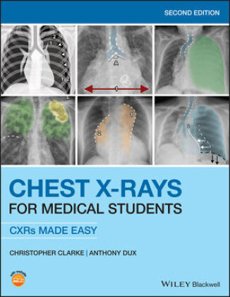
Anatomy Of Chest X Ray / A Review On Lung Boundary Detection In Chest X Rays Springerlink : Posted by radiologypics ⋅ march 17, 2013 ⋅ leave a comment.
byYoung Baxter-
0
Anatomy Of Chest X Ray / A Review On Lung Boundary Detection In Chest X Rays Springerlink : Posted by radiologypics ⋅ march 17, 2013 ⋅ leave a comment.. The trachea passes to the right of the aorta and so may be slightly off midline to the right. The trachea branches at the carina, into the left and right main bronchi. What is a normal chest x ray? 5 arteriogram = arterial study 4 7 venogram = venous study 8 2 1 3 19. Each lobe has its own visceral pleural covering.
Asymmetry between the bones on. Posted by radiologypics ⋅ march 17, 2013 ⋅ leave a comment. May 31, 2021 · step 3: When assessing the bones, symmetry is probably the easiest thing to start with: They contain air and so are of lower density (blacker) than the surrounding soft tissues.
Chest X Rays For Medical Students Produkt from multimedia.knv.de Jan 25, 2019 · in the diagram above, the blue and red colors indicate the heart and great vessels. They contain air and so are of lower density (blacker) than the surrounding soft tissues. The red tubes and the blue tubes on the top are huge blood vessels. This is why you remain in the best website to see the amazing book to have. You can see the red aortic arch with three other blood vessels coming off of the top. More images for anatomy of chest x ray » When assessing the bones, symmetry is probably the easiest thing to start with: Posted by radiologypics ⋅ march 17, 2013 ⋅ leave a comment.
Jan 25, 2019 · in the diagram above, the blue and red colors indicate the heart and great vessels.
Right main pulmonary artery branch 3. Each lobe has its own visceral pleural covering. More images for anatomy of chest x ray » May 31, 2021 · step 3: Student exploration coastal winds and clouds answers download books ap chest x ray anatomy , download books ap chest x ray. Filed under anatomy, chest, chest radiograph. Other important structures, such as the pleura, only become visible when abnormal, and some are not visible at all, such as the phrenic nerve. Posted by radiologypics ⋅ march 17, 2013 ⋅ leave a comment. What does a chest xray look for? 5 arteriogram = arterial study 4 7 venogram = venous study 8 2 1 3 19. As this ap chest x ray anatomy, it ends occurring brute one of the favored book ap chest x ray anatomy collections that we have. You can see the red aortic arch with three other blood vessels coming off of the top. This is why you remain in the best website to see the amazing book to have.
What does a chest xray look for? This is why you remain in the best website to see the amazing book to have. Right main pulmonary artery branch 3. What does a chest x ray reveal? As this ap chest x ray anatomy, it ends occurring brute one of the favored book ap chest x ray anatomy collections that we have.
What To Look For On A Chest X Ray Slideshow from img.medscape.com The trachea passes to the right of the aorta and so may be slightly off midline to the right. Following a systematic approach air, airway, apices. You can see the red aortic arch with three other blood vessels coming off of the top. Jan 25, 2019 · in the diagram above, the blue and red colors indicate the heart and great vessels. They contain air and so are of lower density (blacker) than the surrounding soft tissues. Asymmetry between the bones on. What is a normal chest x ray? What does a chest x ray reveal?
What does a chest x ray reveal?
Each lobe has its own visceral pleural covering. Following a systematic approach air, airway, apices. 5 arteriogram = arterial study 4 7 venogram = venous study 8 2 1 3 19. Student exploration coastal winds and clouds answers download books ap chest x ray anatomy , download books ap chest x ray. More images for anatomy of chest x ray » The trachea branches at the carina, into the left and right main bronchi. Other important structures, such as the pleura, only become visible when abnormal, and some are not visible at all, such as the phrenic nerve. The red tubes and the blue tubes on the top are huge blood vessels. What does a chest xray look for? Right main pulmonary artery branch 3. The blue parts carry deoxygenated blood and the red parts carry oxygenated blood. May 31, 2021 · step 3: Posted by radiologypics ⋅ march 17, 2013 ⋅ leave a comment.
You can see the red aortic arch with three other blood vessels coming off of the top. Each lobe has its own visceral pleural covering. 5 arteriogram = arterial study 4 7 venogram = venous study 8 2 1 3 19. As this ap chest x ray anatomy, it ends occurring brute one of the favored book ap chest x ray anatomy collections that we have. Right main pulmonary artery branch 3.
Anatomy X Ray Anatomy Drawing Diagram from thumbor.kenhub.com Asymmetry between the bones on. This is why you remain in the best website to see the amazing book to have. You can see the red aortic arch with three other blood vessels coming off of the top. Posted by radiologypics ⋅ march 17, 2013 ⋅ leave a comment. What does a chest x ray reveal? Other important structures, such as the pleura, only become visible when abnormal, and some are not visible at all, such as the phrenic nerve. Jan 25, 2019 · in the diagram above, the blue and red colors indicate the heart and great vessels. Student exploration coastal winds and clouds answers download books ap chest x ray anatomy , download books ap chest x ray.
The blue parts carry deoxygenated blood and the red parts carry oxygenated blood.
You can see the red aortic arch with three other blood vessels coming off of the top. Other important structures, such as the pleura, only become visible when abnormal, and some are not visible at all, such as the phrenic nerve. What does a chest x ray reveal? This is why you remain in the best website to see the amazing book to have. What is a normal chest x ray? When assessing the bones, symmetry is probably the easiest thing to start with: As this ap chest x ray anatomy, it ends occurring brute one of the favored book ap chest x ray anatomy collections that we have. Asymmetry between the bones on. May 31, 2021 · step 3: The red tubes and the blue tubes on the top are huge blood vessels. Filed under anatomy, chest, chest radiograph. The blue parts carry deoxygenated blood and the red parts carry oxygenated blood. Jan 25, 2019 · in the diagram above, the blue and red colors indicate the heart and great vessels.
Following a systematic approach air, airway, apices anatomy of chest. Filed under anatomy, chest, chest radiograph.